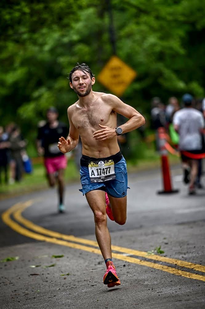
The ACL is a major ligament that connects the thigh bone to the shin bone. While playing sports that require pivoting, such as basketball, the ligament controls sudden stops and changes in direction.
Every female athlete requires a healthy ACL to boost overall performance and participate actively in sports activities.
This blog explains in detail the risk factors, prevention and rehabilitation strategies, ACL injury statistics and return to sport programs on ACL injuries in women.
It can take 9 to 10 months to recover from ACL surgery and return to sports. Women can take 10 months or longer, while men may take 8 months or less to recover.
To determine if you are ready to go back to sports, a physiotherapist ensures that the reconstructed knee is as functional as the other knee.
Females are at a higher risk of contralateral knee injury compared to men. Men have more muscle mass, which may offer protection, while women are at a high risk of re-injury due to hormonal, anatomical and poor landing techniques.
Returning to sports earlier than a 10-month recovery period may put you at risk of reinjury either on the same knee or the opposite knee. This shows that you have not given the new graft enough time to heal.
According to a study done in Australia, the rate at which female athletes returned to sports was lower than that of male athletes, 65% to 75%, respectively.
To prevent reinjury, avoid sports that demand intense jumping, pivoting and quick turns for the first few months.
Following rehabilitation protocol, not returning to sports before you are ready and getting cleared by your physiotherapist is one way to prevent re-injury.
Why Women Are More Prone to ACL Injuries: Key Differences.
As a female athlete, you are likely to tear the ACL(Anterior cruciate ligament ) while participating in sports like soccer, basketball, football and handball. Women tear ACLS more due to ACL injury factors such as Environmental, anatomic, hormonal, and biomechanical factors. Below are ACL injury factors behind higher ACL injury rates in women: Hormonal. The testosterone hormone increases muscle density. The testosterone hormone is more prevalent in men than women; therefore, women's muscles are not able to handle the excess force during sports activities. Women have the estrogen hormone, a hormone responsible for the growth of bones. However, oestrogen hormone fluctuates during periods, leading to loosening of ligaments and tendons. Loosened tendons and ligaments put you at a higher risk of injury. According to research, ACL injuries studied within 24 hours showed that women are at a greater risk of getting ACL injuries during the ovulatory stage, more than the follicular stage. Anatomic. ACL is a less elastic ligament in your knee that connects the femur and tibia. Females have wider hips than men, this changes how the femur, tibia and thigh bone function. Sport games like basketball and soccer require more jumping and pivoting.ACL is responsible for absorbing stress, but when you jump repeatedly, it's difficult for it to absorb too much stress, and may cause a tear. Female has less muscle mass around the knee than males. Less muscle mass puts you at a higher risk of instability and tears due to overstretching of the ligament. Biomechanics. The landing mechanism is the reason why women tear ACl more than men. Safe landing actively engages your core. Female athletes encounter knee ligament vulnerability when landing or pivoting. Female knees buckle inward while landing, therefore increasing the rate of ACL injury. While landing, men land with their knees bent, therefore creating a more core engagement. Another gender difference in sport injuries is quad dominance, where female athletes activate their quadriceps more than their hamstrings when jumping. Imbalance puts more strain on the ACL.Physiological and Hormonal Influences.
Physiological conditions and hormonal influences have a significant impact on ACL. Estrogen and ACL. Estrogen hormone levels are at peak in the pre-ovulatory stage(days 12-14), a period that increases ligament flexibility. High levels of estrogen cause knee instability and likelihood of injury. The estrogen hormone decreases collagen synthesis, and this affects fibre alignment. Ligament laxity weakens the ACL and increases the risk of injury. Fibroblasts on the ACL contain oestrogen receptors that directly impact the laxity of the ligaments and limb alignment. The estrogen hormone plays a significant role during the follicular and ovulatory phases. When the progesterone levels are low, they cannot inhibit the effects of estrogen on the ACL. Hormonal impact on ligaments. Hormonal fluctuations of hormones such as relaxin increase the risk of ACL injury. Females have a higher concentration of relaxin hormone during the luteal phase of the period. High relaxin hormone increases ACL laxity, therefore putting you at risk of getting a rupture. Not enough research shows that the oral contraceptive pill reduces hormonal fluctuation and laxity of the ACL during the menstrual cycle.Body Composition and Muscle Distribution.
ACL injuries are higher in women compared to men due to differences in body composition and muscle distribution. Muscle strength and flexibility impact ACL. Imbalanced muscles in the hamstrings and quadriceps cause improper knee mechanics and strain your ligaments hamstrings and quadriceps stabilise knee joints and control knee movements. Women have lower muscle mass in the lower body than men. During high-impact movements such as cutting or jumping, the lower body cannot handle the force placed on the ACL.Skeletal structure and Pelvic Length.
Pelvic width ACL risk is higher in females compared to men. The wider the pelvic floor, the greater the quadriceps angle (Q angle). When the Q angle is greater than 19 degrees, you are at risk of getting Q-angle injuries. Q angle affects knee alignment and how weight is distributed on the knee joint. When you participate in high-impact movements such as jumping, pivoting and cutting, you place more strain on the ACL. Female knees tend to bend inwards, spreading more weight and force to the lateral side of the knee, therefore stressing the ACL. Too much force weakens ligaments and reduces the strength and stability of your knee. Stress can also lead to ligament tears that may affect your participation in sports activities. Q angle in women affects the angle at which the quadriceps muscles connect with the patella and the direction in which the patella is pulled. A greater Q angle means that the patella will be moved laterally, whereby the lateral pull increases as flexion increases, and this can lead to dislocation of the patella. Female athletes' body composition has a narrower intercondylar notch compared to men. The intercondylar notch is the space in the knee where the ACL is positioned. The narrower the intercondylar, the higher your risk of tearing your ACL during high-impact knee movements.Biomechanical Risk Factors.
Plant and cut, rotating and landing from a jump are movements that you don't plan for. These movements happen as a form of reaction to a ball or defending against a score. Cutting and decelerating are non-contact ACL injuries that soccer players frequently execute.ACL injuries are likely to occur while playing handball due to plant and cut movements or landing from a jump. Poor landing mechanics put you at risk of ACL injury. 70% of the time spent playing basketball puts you at risk of ACL injuries due to high-risk movements and poor landing mechanisms. There is a difference in how the knee rotates inwards and outwards while side-step cutting for men and women. Females experience too much inwards rotation of the tibia than the femur, and this overstretches the ACL, increasing the chances of an injury. Female athletes land with their hips bent(increased hip flexion) and sustain the position for longer periods compared to males. Sustaining the hip flexion position for long periods puts female athletes at a risk of non-contact ACL injury. During vertical jumps while participating in sports like handball and football, females with ACL injuries land quicker and experience more knee abduction.Key Anatomical Concepts.
The ACL is located in the knee joint, connecting the femur and tibia.ACL is made up of (AMB) anteromedial and (PLB) posterolateral. The PLB fascicle originates from the distal part of the femoral attachment, while the AMB originates from the posterior part of the femoral attachment. When you bend the knee, the AMB lengthen and tightens while the PLB shorten and becomes loose. When you extend your knee, the PLB lengthen and tightens while the PLB remain tight but not tighter than AMB. The main role of ACL is to stabilise the knee joint. It stabilises the anterior tibial translation and resists internal rotation. It also prevents excessive movements during sports activities. When you extend your knee, the anterior tibial translation is at 2mm maximum and can support your knee while standing. When you bend your knee, the anterior tibial translation increases to 3mm when walking and increases to 6mm when force is applied at the anterior position. If your ACL is damaged, the anterior tibial translation can increase up to 15mm at 30 degrees flexion when force is applied. Tibia slope determines the position of the tibia to the femur. Females with ACL injuries have greater tibial slopes compared to men with ACL injuries. Increased posterior tibial slope is a risk factor for ACL injury.ACL Injury Statistics in Female Athletes.
Globally, women suffer ACL injuries more than male athletes. Below are statistics of female ACL tear rates compared in various sports: In 1000-athlete exposures, females are about three times more likely to get ACL injuries than men. Player position and player movement are key factors in an ACL injury.| Sport | Male injury statistics | Female injury statistics |
| Volleyball | 1 | 3 |
| Soccer | 1 | 4 |
| World Cup skiing | 1 | 3 |
| Basket ball | 1 | 3.5 |
Comprehensive Assessment.
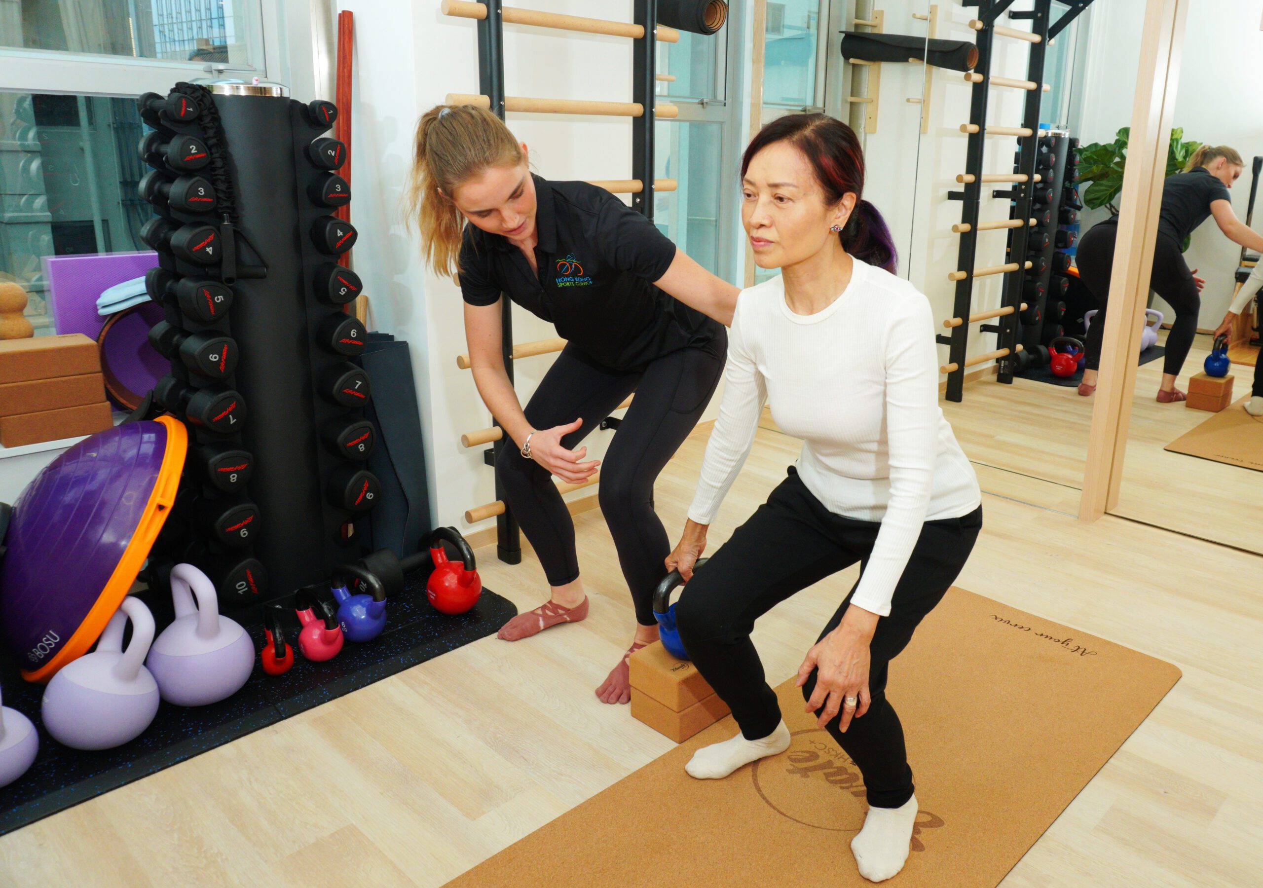
At Hong Kong Sport Clinic, we do gait analysis where we look from the front, back and side of your leg for ACL injury assessment. During gait, we do injury diagnostics where we assess certain aspects of your legs. A gait cycle is included in this assessment. In physiotherapy evaluation, a physiotherapist assesses your muscle strength to get information about neurological deficits. Your healthcare provider will be able to differentiate if it's a weakness or an imbalance. In Hong Kong, we do sport-specific screening that involves screening your physical condition, body fitness, and other potential health risks that may prevent you from participating in sports activities. Our flexibility tests focus on evaluating your range of motion, ability to stretch and joint flexibility. Flexibility tests include both laboratory and field testsCustomised Rehabilitation Protocol.
ACL rehab phases help in physiotherapy intervention and are divided into the following phases: Acute Stage. The acute stage focuses on improving range of motion, strength and stability. Your physiotherapist will recommend exercises to strengthen the quadriceps and hamstrings, range of motion and proprioception. Anti-inflammatory treatments help in reducing swelling and pain. Get the right anti-inflammatory prescriptions from your doctor to help control pain and swelling. In the acute and sub-acute phase, your physiotherapist may encourage:- Achieving full extension through heel props, static quads and passive knee extension.
- Enhance bending through wall slide, heel slides and passive knee bend.
- Weight transfers in standing
- Knee bending and extension in sitting
- Swimming.
- Squats
- Lunges
- Leg press machine
- Step-ups.
- One leg standing position in the frontal and sagittal planes.
- One-leg standing exercises.
- One leg standing exercises in combination with balance cushions
- Regain the full strength of your lower limb.
- Ability to hop perefectly
Return-to-Sport Programming.
Returning to sport after ACL should not be done on assumption, however, factors like strength benchmarks, psychological readiness, sport-specific drills and injury prevention procedures are critical. Sport-specific drills mimic the physical activities of specific sports. Examples of sport-specific drills include:- Plyometric exercises enhance neuromuscular control. This type of exercise should be done by female basketball, football and soccer athletes to increase knee flexion angle and reduce knee valgus during a drop vertical jump. An example of plyometric exercises is jump training.
- Progressive Agility training mimics specific sports activities. It includes movements such as pivoting, jumping, and cutting. This training improves coordination so that you can perform the movements safely.
- Unanticipated movement drills improve coordination, strength, and timing, ensuring that your quadriceps and hamstrings work together to stabilise your knee.
- Modifying your techniques. Practising proper landing mechanics and cutting techniques reduces knee valgus. A physiotherapist may help identify improper forms and suggest correct techniques.
- Neuromuscular training programs improve coordination, balance and strength. If young female athletes are introduced to neuromuscular training before puberty, they are less likely to be at risk of ACL injury.